Ganglioneuroma no Mediastino Posterior Identificado após Investigação de Tosse Persistente em Criança: Relato de Caso
Barra lateral de artigos
Conteúdo do artigo principal
Resumo
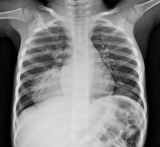
O ganglioneuroma (GN) é um tumor neuroblástico, que se origina a partir de células da crista neural. São raros, benignos e frequentemente assintomáticos, apresentando sintomatologia quando há compressão de estruturas adjacentes. Neste relato, os autores apresentam um caso de tosse persistente em um menino de três anos de idade com um tumor torácico extenso, cujo diagnóstico foi de ganglioneuroma no mediastino posterior, focando os aspectos mais importantes do diagnóstico desta patologia. Este caso destaca a importância da investigação por imagem em quadros pediátricos respiratórios refratários, alertando para a inclusão de tumores neuroblásticos no diagnóstico diferencial.
Detalhes do artigo

Este trabalho está licenciado sob uma licença Creative Commons Attribution 4.0 International License.
Authors retain the copyright of their articles and grant the journal the right of first publication under the Creative Commons Attribution (CC BY) license, which allows others to share and adapt the work with proper attribution.
Referências
Perdomo Reyes C, Chambon C, Gonzalez Gonzalez D. Ganglioneuroma suprarrenal: reporte de un caso. Rev Med Urug. 2020 Aug 1;36(3).
Silva J, Cachulo MC, Leitão-Marques A. Establishing a secure connection … [Internet]. SciELO Brasil. 2025 [cited 2025 Oct 7]. Available from: https://www.scielo.br/j/abc/a/7nwhc4NscCq8SQxGVXRWQQP/?lang=en.
Ferreira Oliveira A, José Vieira L, Alexandre Moreira A, Baptista de Paula Fraga J, Ribeiro Lourenço Costa R. Ganglioneuroma retroperitoneal: relato de caso. Ganglioneuroma retroperitoneal: case report [Internet]. [cited 2025 Oct 7]. Available from: https://cdn.publisher.gn1.link/relatosdocbc.org.br/pdf/n1a07.pdf.
Guan YB, Zhang WD, Zeng QS, Chen GQ, He JX. CT and MRI findings of thoracic ganglioneuroma. Br J Radiol. 2012 May 10;85(1016):e365–72.
Majbar A, Elmouhadi S, Elaloui M, Raiss M, Sabbah F, Hrora A, et al. Imaging features of adrenal ganglioneuroma: a case report. BMC Res Notes [Internet]. 2014 [cited 2025 Oct 7];7:791. Available from: https://pmc.ncbi.nlm.nih.gov/articles/PMC4289252/.
Pacella G, Brunese MC, Donnarumma F, Barrassi M, Bellifemine F, Sciaudone G, et al. Imaging of ganglioneuroma: a literature review and a rare case of cystic presentation in an adolescent girl. Diagnostics (Basel) [Internet]. 2023 Jan 1 [cited 2023 Oct 6];13(13):2190. Available from: https://www.mdpi.com/2075-4418/13/13/2190.
Rueda-de-Eusebio A, de la Torre Serrano M, Victoria Artalejo A, Mendez R. Adrenal ganglioneuroma with radiology–pathology correlation. Cureus [Internet]. 2024 Sep 17 [cited 2025 Oct 7]. Available from: https://www.cureus.com/articles/281594-adrenal-ganglioneuroma-with-radiology-pathology-correlation.
Zhang QW, Song T, Yang PP, Hao Q. Retroperitoneum ganglioneuroma: imaging features and surgical outcomes of 35 cases at a Chinese institution. BMC Med Imaging. 2021 Jul 22;21(1).
Gahr N, Darge K, Hahn G, Kreher BW, von Buiren M, Uhl M. Diffusion-weighted MRI for differentiation of neuroblastoma and ganglioneuroblastoma/ganglioneuroma. Eur J Radiol [Internet]. 2010 May 13;79(3):443–6. Available from: https://www.sciencedirect.com/science/article/pii/S0720048X1000149X.
Shin JH, Lee HK, Khang SK, Kim DW, Jeong AK, Ahn KJ, et al. Neuronal tumors of the central nervous system: radiologic findings and pathologic correlation. Radiographics. 2002 Sep;22(5):1177–89.
Lasca A, Laia I, Pires Santos R, Dias Carneiro A, Moreira D. Paravertebral ganglioneuroma in pediatric age: a case report. Cureus. 2024 Jun 28.
Lonergan GJ, Schwab CM, Suarez ES, Carlson CL. From the archives of the AFIP. Radiographics. 2002 Jul;22(4):911–34.