Surgical Management of Submandibular Gland Sialolithiasis: A Case Report
Article Sidebar
Main Article Content
Abstract
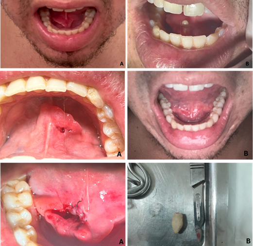
Sialolithiasis is an inflammation caused by the obstruction of a salivary gland’s excretory system due to the formation of calculi. These mineralized structures occur mainly in the major salivary glands, particularly in the submandibular gland, due to the higher viscosity of saliva and increased concentrations of calcium and phosphate. The formation of sialoliths involves factors such as low salivary flow, infections, dehydration, and pH alterations, which lead to solute crystallization. Treatment aims to remove the sialolith; when the calculus is small and located near the duct opening, duct dilation and gland massage may result in its expulsion. However, if these conditions are not met, surgical removal becomes necessary. This case report describes a 26-year-old patient who presented to an Oral Surgery service with persistent pain in the submandibular region caused by a sialolith. Despite the absence of radiographic examination, the presence of a calculus in the submandibular duct was confirmed clinically. Surgery was performed under local anesthesia, resulting in successful removal of the sialolith. Postoperative medication and care instructions were provided. Despite the prolonged nature of the condition, the treatment yielded satisfactory results, with no signs of complications or recurrence.
Article Details

This work is licensed under a Creative Commons Attribution 4.0 International License.
Authors retain the copyright of their articles and grant the journal the right of first publication under the Creative Commons Attribution (CC BY) license, which allows others to share and adapt the work with proper attribution.
References
Gulati U, Singh S, Kaur R, Kaur H. Submandibular sialolithiasis: a brief overview and report of two cases. Modern Res Dent. 2018;1(5):1-7.
Holden AM, Adamson R, Davies-Husband C, Bentley R. Audit of minimally-invasive surgery for submandibular sialolit-hiasis. Br J Oral Maxillofac Surg. 2019;57(6):582-586.
Kolomiets SV, Melnichenko YM, Ivanova EV, Kravtsova OA. Difficulties in diagnosis of sialolithiasis: A case series. Bull Tokyo Dent Coll. 2018;59(1):53-58.
Sherman JA, McGurk M. Lack of correlation between water hardness and salivary calculi in England. Br J Oral Maxillofac Surg. 2000;38(1):50-53.
Diebold S, Overbeck M. Soft Tissue Disorders of the Mouth. Emerg Med Clin North Am. 2019;37(1):55-68.
Marchal F, Dulguerov P. Sialolithiasis management: the state of the art. Arch Otolaryngol Head Neck Surg. 2003;129(9):951-956.
Arifa SP, Muhammed S, Saleem S, Ravi MB. Sialolithiasis of the Submandibular Gland: Report of Cases. Cureus. 2019;11(3):4180.
Oliveira TP, Silva LF, Martins-Filho PR, Santos TS. Giant sialolith of submandibular gland duct treated by excision and ductal repair: a case report. Braz J Otorhinolaryngol. 2016;82(1):112-115.
Folchini S, Stolz AB. Sialoliths in the submandibular gland: a case report. Odontol Clin-cient. 2016;15(1):67-71.
Aisshwarya P, Sivapathasundharam B, Santhosh Kumar MP. Diagnosis and management of submandibular duct sialoliths: Report of 2 cases. Nac J Med Den Res. 2017;5(3):237-241.
Da Silva WG, de Souza Andrade ESS, de Oliveira Costa F, de Souza Figueiredo PT. Stafne’s bone defect in a metastatic prostate cancer patient: A diagnostic conundrum. J Clin Exp Dent. 2018;10(1):88-91.
Huoh KC, Eisele DW. Etiologic factors in sialolithiasis. Otolaryngol Head Neck Surg. 2011;145(6):935-939. doi: 10.1177/0194599811415801.
Iqbal A, Gupta AK, Natu SS, Gupta AK. Unusually large sialolith of Wharton's duct. Ann Maxillofac Surg. 2012;2(1):70-73.
Rzymska-Grala I, Stopa Z, Grala B, et al. Salivary gland calculi - contemporary methods of imaging. Pol J Radiol. 2010;75(3):25-37.